Case 2 presentation
contributed by
Dr. Detlef Engineer and Dr. Katja Kirchhoff, Augenklinik Tausendfensterhaus, Duisburg, Germany
Categories: Vitreoretinal, vascular changes
Key problem: Finding of unknown etiology with necessity of urgent treatment
In contrast to other case reports, we present expert-level cases without an unambiguous solution: A case report with a solution will only add more encyclopedic knowledge to Your repertoire, while an open-ended case report will train Your methods. Therefore each case highlights a specific problem expressed in the introduction.We won’t offer the satisfying feeling of solving a puzzle correctly, but something more valuable: You will train Your skills by developing proper methods for “how to proceed” and You will help others to train their skills by giving them your perspective.Don´t be afraid to send Your opinion even if You do not feel safe. It’s meant to be tough and You may stay anonymous, if You wish so.
Female, 46 years old, Asian
History of sudden vision loss left eye 6 weeks prior to consultation in our clinic. Patient states that vision was full on both eyes prior to incident. Examinations have been made at home (in China) directly after event. Patient brings print-outs of fundus photography, angiography, OCT and sonography.
Best-corrected visual acuity: OD 20/20, OS hand motion
IOD: 15/12 mmHg
Anterior segment: OD within normal limits, OS severe vitreous hemorrhage, otherwise within normal limits
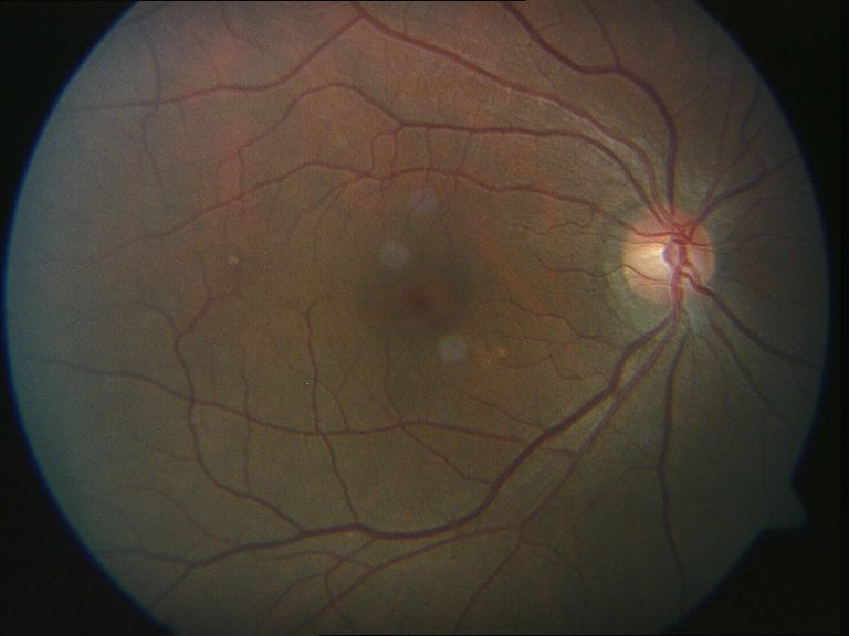
Fundus: OD no abnormalities detected, OS not visible because of vitreous hemorrhage
Plan: OS vitrectomy performed
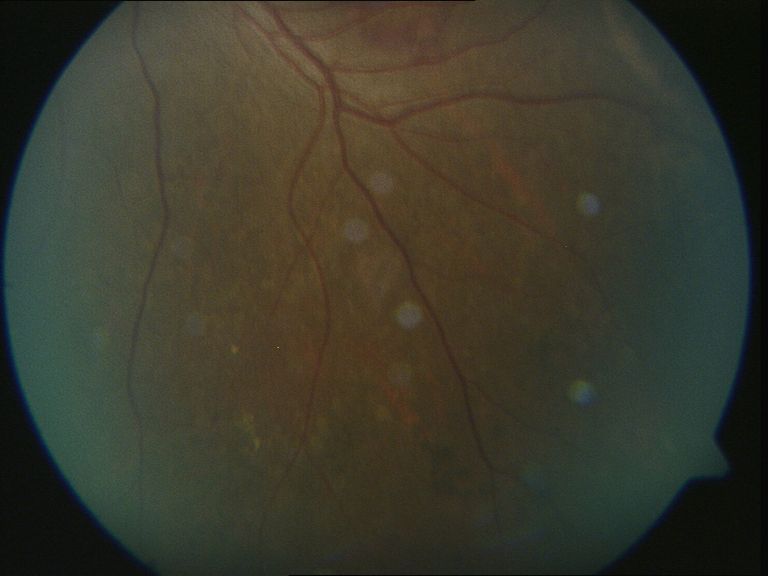
Fundus (print-outs brought by patient)

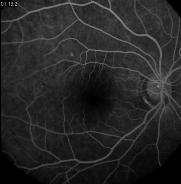
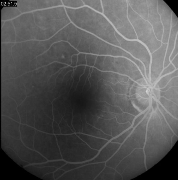
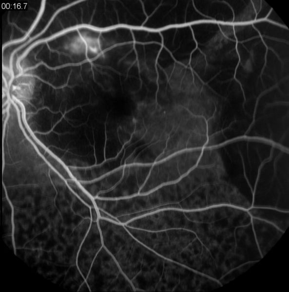
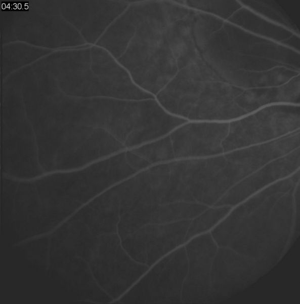
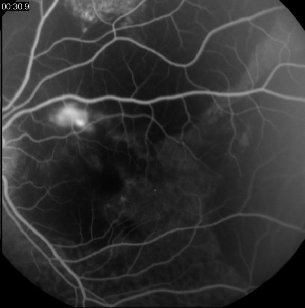
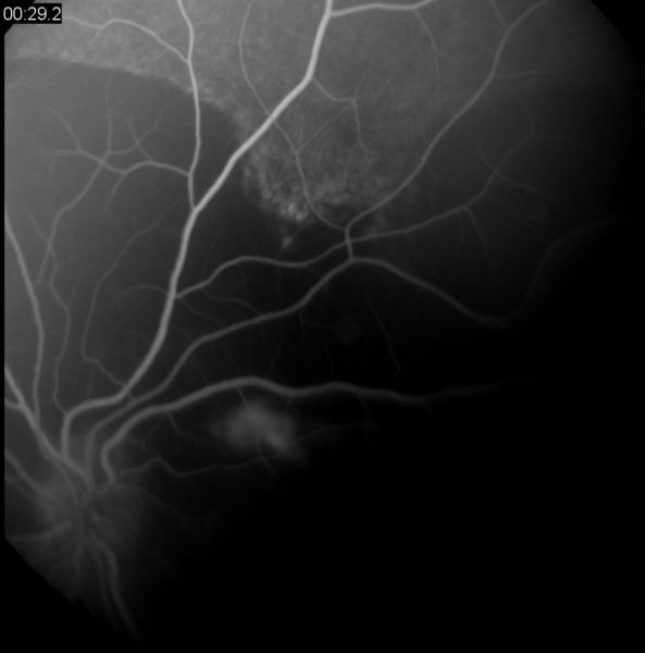
Angiography (print-outs brought by patient)
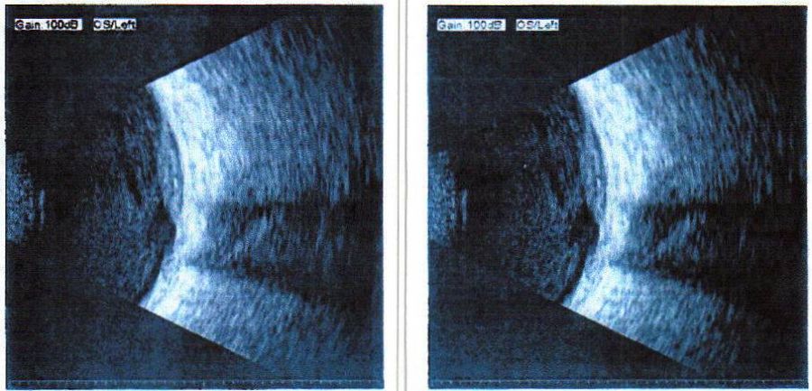
Sonography (print-outs brought by patient)
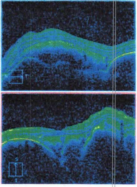
OCT (print-outs brought by patient)
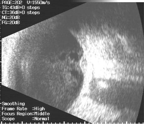
Sonography (on examination)
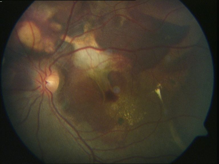
Fundus (after vitrectomy)
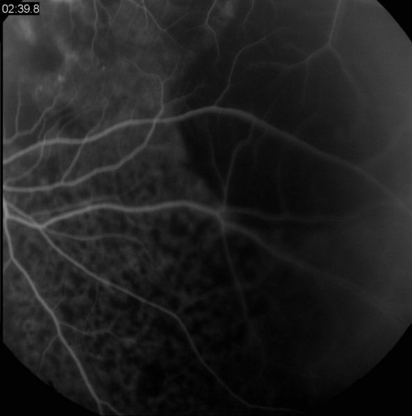
Angiography OD (after vitrectomy)
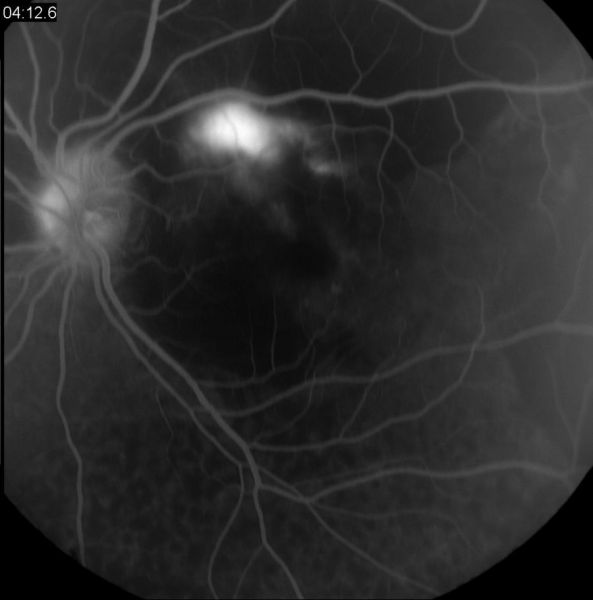
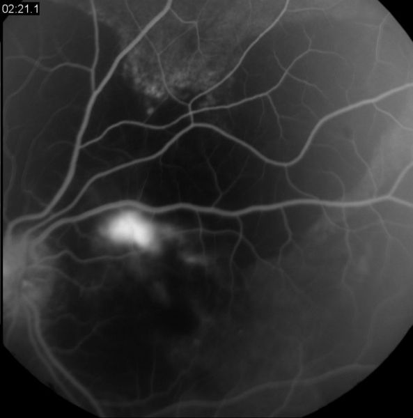
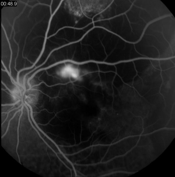
Angiography OS (after vitrectomy)
What might have caused these findings and how would You have proceeded?
This is a “hidden” case – You have to send Your opinion first to see others´ opinions


