


Case 3 presentation
contributed by
PD Dr. Tobias Stupp, Meerbusch, Germany and Dr. Philipp Franko Zeitz, Düsseldorf, Germany
Categories: Cornea, infections, herpetic disease, surface disorders
Key problem: Multiple different findings that need treatment, but the therapy for one will worsen the other
In contrast to other case reports, we present expert-level cases without an unambiguous solution: A case report with a solution will only add more encyclopedic knowledge to Your repertoire, while an open-ended case report will train Your methods. Therefore each case highlights a specific problem expressed in the introduction.We won’t offer the satisfying feeling of solving a puzzle correctly, but something more valuable: You will train Your skills by developing proper methods for “how to proceed” and You will help others to train their skills by giving them your perspective.
Don´t be afraid to send Your opinion even if You do not feel safe. It’s meant to be tough and You may stay anonymous, if You wish so.
Female, 65 years old
History of metastasized ovarian carcinoma with recent chemotherapy (gemcitabine, carboplatin, bevacizumab, premedication: ondansetron, aprepitant, ranitidine, dexamethasone)
First examination (day 1)
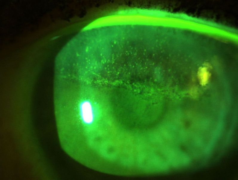
Anterior segment: OS herpetic keratitis with multiple dendritis ulcerations (see photo), otherwise within normal limits
Therapy: Aciclovir ointment five times a day

Second examination (4th week)
Anterior segment: OS good improvement of the herpetic alterations with no more dendritic ulcerations visible, but now remarkable inhomogenous opacities in the upper cornea with dotted fluoresceine staining and a demarcation line with fluoresceine pooling at the inferior edge (see photo)
Therapy: Aciclovir stopped, artificial tears every two hours
Third examination (12th week)
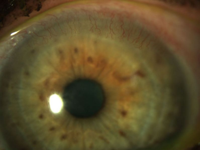
Anterior segment: OS unchanged on fluoresceine staining, but now progressive corneal vascularization (see photo)
How would You proceed at this point?

